|
Summary
| Background
| Methods | Results | Publications
|
Microtubules and Their Plus-ends
|
|
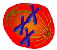 Microtubules
are long, tube-shaped (25 nm in diameter) protein polymers that are present
in all eukaryotic cells. An important function of microtubules is to drive
the movement of cellular organelles such as chromosomes. During mitosis,
microtubules attach their end structures, called plus-ends in this case, to
chromosomes via kinetochore (a secondary structure of chromosomes),
illustrated in the left figure. As the microtubules grow and shrink, the
chromosomes are pulled around inside the cell. Because of their vital roles
in cells, microtubules have been under intensive investigation in both cell
biology and molecular medicine. For example, a direction in anti-cancer
drug development is to disrupt microtubule dynamics to stop cells from
proliferating. Microtubules
are long, tube-shaped (25 nm in diameter) protein polymers that are present
in all eukaryotic cells. An important function of microtubules is to drive
the movement of cellular organelles such as chromosomes. During mitosis,
microtubules attach their end structures, called plus-ends in this case, to
chromosomes via kinetochore (a secondary structure of chromosomes),
illustrated in the left figure. As the microtubules grow and shrink, the
chromosomes are pulled around inside the cell. Because of their vital roles
in cells, microtubules have been under intensive investigation in both cell
biology and molecular medicine. For example, a direction in anti-cancer
drug development is to disrupt microtubule dynamics to stop cells from
proliferating.
|
|
Problems and Challenges
|
|
|
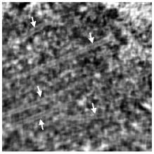
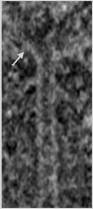
Biologists
study the functions of microtubules and plus-ends by observing their
structures. For this purpose, electron microscope tomography is used to produce
high-resolution 3D volume image. However, in the low contrast tomography
volume, the interpretation of the volume data is rather challenging since
the microtubules and plus-end features are in close contact with the
cellular environment and are densely surrounded by proteins of similar
appearance, as shown in both the left and right figures. Consequently,
practical segmentation of the tomography volume is dominated by
time-consuming manual operation.
|
|
|
A selected slice in the volume, microtubules are
indicated by arrows
|
A close-up view of one microtubule, plus-end indicated by arrow
|
Summary | Background | Methods | Results | Publications
|